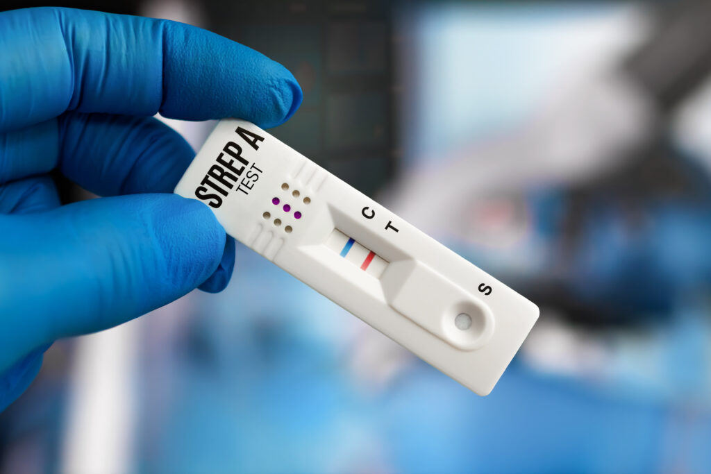
Group A Streptococcus (Streptococcus pyogenes) is an aerobic gram-positive coccus that causes many infections. GAS is most commonly associated with pharyngitis or skin and soft tissue (non-necrotizing) infection; these are not typically associated with invasive infection.
Less commonly, GAS causes invasive disease; invasive GAS infection refers to infection in the setting of culture isolation of GAS from a normally sterile site (most commonly blood; less commonly pleural, pericardial, joint, or cerebrospinal fluid).
- Forms of invasive GAS infection include:
- Necrotizing soft tissue infection
- Pregnancy-associated infection)
- Bacteremia (may occur in association with or in the absence of another infection)
- Respiratory tract infection
Toxic shock syndrome (TSS) occurs as a complication of invasive GAS disease in approximately one-third of cases- See TSS article.

Invasive infections associated with GAS have been reported with increasing frequency, predominantly from North America and Europe.
Epidemiology
Incidence — In resource-rich settings, there are an estimated 3.5 cases of invasive GAS infection per 100,000 persons, with a case-fatality rate of 30 to 60 percent.
Invasive infections associated with GAS have been reported with increasing frequency, predominantly from North America and Europe. In the United States, invasive GAS infections increased in the late 1980s. After that, the incidence was stable from the mid-1990s until 2012 but has increased subsequently (average annual rate of 3.8 per 100,000 from 2005 to 2012; rate of 4.8 to 5.8 per 100,000 for 2014 to 2016).
Approximately 100 to 200 pediatric cases were reported to the Active Bacterial Core (ABC) surveillance at the Centers for Disease Control and Prevention (CDC) each year from 2015 through 2019. There were only 74 cases reported in 2020, likely due to the impact of social distancing and other infection control measures during the height of the COVID-19 pandemic. Similarly, in a Texas Children’s Hospital report, there were considerably fewer hospital admissions for invasive GAS infections in 2020 compared with previous years, though the rate increased again by 2022 . In December of 2022, the CDC issued a health advisory regarding an increase in pediatric invasive GAS infection cases reported to the ABC surveillance program in November through early December.
In general, invasive GAS infection may occur in patients of any age. The incidence is highest in adults >50 years of age, followed by young children (particularly those <1 year of age). Most patients are not immunosuppressed. Since the 1980s, an increase in the frequency of invasive GAS disease has occurred among patients 20 to 35 years of age. This change likely reflects the increased frequency of two presentations of infection: (1) pregnancy-associated GAS infections and (2) deep infection at the site of muscle injury or blunt trauma in the absence of a clear portal of entry .
The factors responsible for the increase in the incidence of invasive GAS infection are not fully understood. Alterations in the distribution of GAS strain M types and an increasing prevalence of toxin-producing strains may be responsible. About half of the invasive GAS infections are caused by a limited number of GAS M types (1, 3, 4, 6, and 28); the remaining half is caused by various strains, including non-typeable strains. All GAS strains isolated from invasive infections produce a toxin called NADase.
Risk Factors
Risk factors associated with the development of invasive GAS infection include:
- Minor trauma, including injuries resulting in hematoma, bruising, or muscle strain
- Use of nonsteroidal anti-inflammatory drugs (NSAIDs)
- Recent surgery
- HIV infection
- Other viral infections (eg, influenza, varicella)
- Intravenous drug use
- Burns
- Obesity
- Malignancy
- Corticosteroid use
- Diabetes mellitus and other forms of immunosuppression
- Influenza Infection
Risk factors for progression from invasive GAS infection to TSS are poorly defined. Failure to recognize the condition (resulting in delays in antibiotic administration and surgical debridement) is a potentially important risk factor. In addition, it has been suggested that major histocompatibility complex class II haplotypes may influence host susceptibility to the development of TSS .
Invasive GAS Infection (In Abscence of Toxic Shock)
The most common portals of GAS entry are the skin, vagina, and pharynx.
Invasive GAS disease forms include necrotizing soft tissue infection, pregnancy-associated infection, bacteremia, and pneumonia.
Necrotizing soft tissue infection — Necrotizing soft tissue infection may involve the epidermis, dermis, subcutaneous tissue, fascia, and muscles. Necrotizing infection most commonly involves the extremities. It can be predisposed in children by cellulitis, minor trauma, burns, and varicella-zoster virus (VZV) infection most commonly. Affected patients may have other signs of invasive GAS infection, such as osteomyelitis, septic arthritis, necrotizing fasciitis, or myonecrosis. Necrotizing infection usually presents acutely (over hours); it rarely presents subacutely (over days). Rapid progression to extensive destruction can occur, leading to systemic toxicity, limb loss, and/or death.
GAS is one of several causes of necrotizing skin infections, but this is the classic syndrome of invasive GAS infection. TSS occurs as a complication of necrotizing soft tissue infection in approximately half of cases.
Pregnancy-associated infection — Invasive GAS infection associated with pregnancy (also referred to as puerperal GAS sepsis or postpartum GAS infection) is a significant cause of maternal and infant mortality.
Most cases of puerperal GAS sepsis occur during the first 48 to 72 hours after delivery. In approximately 20 percent of cases, invasive GAS infection occurs during late pregnancy (prior to onset of labor or rupture of membranes). Invasive GAS infection can also occur after discharge from the hospital. Patients with puerperal sepsis due to GAS typically present with fever and abdominal pain.
Postpartum endometritis can be caused by a number of organisms, including GAS; GAS endometritis can progress to puerperal sepsis. Postpartum endometritis should be suspected in the setting of fever and uterine tenderness following childbirth.
Bacteremia — GAS bacteremia usually occurs associated with infection at a primary site [40,41].
- The most common source of GAS bacteremia is skin and soft tissue infection; forms include cellulitis/erysipelas, surgical wound infection, varicella virus infection, and burns
- GAS bacteremia may also occur in the setting of necrotizing soft tissue infection, pregnancy-associated infection, or respiratory tract infection.
In some cases, GAS bacteremia occurs in the absence of a clear localizing source. Symptoms may be nonspecific and include fever, chills, and fatigue. Among older patients, diabetes and peripheral vascular disease may serve as predisposing factors for isolated GAS bacteremia. Additional risk factors (among children and older adults) include malignancy and immunosuppression.
Less common manifestations — Uncommonly, GAS can cause pneumonia; in some cases, multilobular diffuse disease may develop. Pleural effusion (particularly left-sided effusion) can also occur; when present, pleural effusion associated with GAS pneumonia often represents an empyema. Among adults with pneumonia due to GAS, bacteremia occurs in approximately 80 percent of cases.
GAS bacteremia has been associated with suppurative complications of pharyngitis such as peritonsillar abscess and with extension of infection into the sinuses, middle ear, and mastoids.
Other (relatively uncommon) manifestations of invasive GAS infection include septic arthritis, osteomyelitis, peritonitis, and meningitis.
Diagnosis — Invasive streptococcal infection should be suspected in patients with signs of systemic illness (such as fever) in the setting of skin or soft tissue infection; it should also be suspected in pregnant and postpartum women in the setting of high fever or rapid onset of fever. GAS bacteremia may occur in the absence of a clear localizing source.
The diagnosis of invasive GAS infection is established via positive culture for GAS from a normally sterile site (most commonly blood; less commonly pleural, pericardial, joint, or cerebrospinal fluid).
For patients with suspected invasive GAS infection, blood cultures should be obtained (ideally prior to antibiotic administration). In addition, cultures should be collected from clinically relevant sites. For patients with skin or soft tissue infections, a wound culture should be obtained. Patients who undergo debridement should have debrided material sent for culture. Postpartum women should have endometrial aspiration for Gram stain and culture. Patients with pneumonia should have throat and sputum culture; in addition, patients who undergo thoracentesis should have pleural fluid sent for culture and pleural fluid chemistries. (See Recovery of GAS from blood cultures usually takes 8 to 24 hours. Gram stain of involved tissue demonstrating gram-positive cocci in pairs and chains can provide an early diagnostic clue in many cases.
Treatment
Susceptibility testing should be pursued for clindamycin and oxazolidinones (linezolid and tedizolid), given the importance of these agents in suppressing toxin production.
References
Tapiainen T, Launonen S, Renko M, et al. Invasive Group A Streptococcal Infections in Children: A Nationwide Survey in Finland. Pediatr Infect Dis J 2016; 35:123.
Nelson GE, Pondo T, Toews KA, et al. Epidemiology of Invasive Group A Streptococcal Infections in the United States, 2005-2012. Clin Infect Dis 2016; 63:478.
Centers for Disease Control and Prevention. Active Bacterial Core Surveillance (ABCs). ABCs Report: group A Streptococcus, 2019. Available at: https://www.cdc.gov/abcs/downloads/GAS_Surveillance_Report_2019.pdf (Accessed on December 03, 2021).
Tyrrell GJ, Lovgren M, Kress B, Grimsrud K. Varicella-associated invasive group A streptococcal disease in Alberta, Canada–2000-2002. Clin Infect Dis 2005; 40:1055.
Carapetis JR, Jacoby P, Carville K, et al. Effectiveness of clindamycin and intravenous immunoglobulin, and risk of disease in contacts, in invasive group a streptococcal infections. Clin Infect Dis 2014; 59:358.
Jaggi P, Beall B, Rippe J, et al. Macrolide resistance and emm type distribution of invasive pediatric group A streptococcal isolates: three-year prospective surveillance from a children’s hospital. Pediatr Infect Dis J 2007; 26:253.
